Watching a patient walk is one of the oldest tools in the clinical exam, and one of the least standardized. A clinician who's been practicing 20 years might catch a subtle subtalar drift in three steps. The new graduate next door might miss it entirely. Both are doing “gait analysis.” Both call it clinical reasoning. Neither is wrong, exactly. But the peer-reviewed literature on observational gait analysis is sobering, and worth a careful read for any practice that prescribes off the back of a visual exam.
This post walks through what the research actually says about visual gait scoring, where instrumented data adds rigor, and how foot capture, the part many practices have historically delegated to a lab, fits into a defensible orthotic prescription. Written for the providers who use gait in their exams, in chiropractic, physical therapy, podiatry, and pedorthic practices alike, and the staff who run them.
What the literature says about observational gait analysis
Observational gait analysis (OGA), watching a patient walk and scoring what you see, has been studied for decades. The pattern across the research is consistent: reliability is moderate at best, even with experienced clinicians and standardized scoring instruments.
A 2025 review in the Journal of Physical Therapy Science examined the reliability and validity of observational gait analysis by physical therapists and concluded that visual joint-angle estimation produced inter-rater reliability that ranged broadly across raters and joints, with experience helping but not eliminating the gap. A separate systematic review on stroke patients found that even when standardized tools (the Hemiplegic Gait Analysis form, Rivermead Visual Gait Assessment, Gait Assessment and Intervention Tool, Wisconsin Gait Scale) were used, measurement properties varied considerably across instruments.
The gist: visual gait scoring is useful as a clinical screen and an exam-room conversation starter. It is not a measurement-grade input. Treating it as one is where over-confident prescribing tends to start.
Visual gait analysis is a clinical screen, not a measurement. The literature has been clear on this for thirty years.
Why this matters for orthotic prescribing
The downstream effect of moderate-reliability inputs is moderate-reliability outputs. If two clinicians watching the same patient disagree on the rearfoot pattern they're seeing, the orthotic prescriptions that flow from those observations will diverge too. Multiply that across a busy practice prescribing dozens of pairs a month, and you get a quiet but real source of remake-rate variance.
This isn't an argument against observational gait analysis. It's an argument for layering it with capture data that doesn't depend on the observer.
The kinetic-chain story
The clinical case for gait-aware orthotic prescribing is well-established, and chiropractic has argued it longer than most. The foot-spine connection, how mechanical asymmetries at ground contact propagate up the kinetic chain to the pelvis, lumbar spine, and beyond, is a core part of how chiropractic frames orthotic intervention, and the same logic drives gait work in physical therapy, podiatry, and pedorthics. Foot Levelers built a multi-decade business around this argument, and the chiropractic literature broadly supports the kinetic-chain framing as a clinical model worth taking seriously.
What the research is less generous about is the specific claim that visual gait observation alone provides enough information to prescribe a custom orthotic. The orthotic itself is a millimeter-grade device. The input that drives it deserves to be a millimeter-grade measurement.
Where instrumented capture adds rigor
Instrumented gait analysis (force plates, 3D motion capture, plantar pressure mapping) is the gold standard in research settings. It also produces output the clinician can defend in writing if a remake gets challenged or a payer asks for documentation.
For most clinical practices, full motion-capture systems are overkill (the labs that have them tend to be university-affiliated). What's reasonable in a clinical practice is a tighter version of the same idea: pair the visual exam with an instrumented foot capture that produces a reproducible 3D digital cast you can refer back to. That's the input the prescription is built on.
What the research says about foot scanners
Here the literature has evolved meaningfully in the last few years. A 2025 study in Sensors examined whether scanner choice matters for the design of foot orthoses and found that different 3D scanners captured similar gross foot morphology, but differed in their accuracy on the small-detail measurements, the millimeter-level features that orthotic prescriptions actually hinge on. A 2020 systematic review in the Journal of Foot and Ankle Research compared 3D scanning to traditional plaster casting and found 3D capture broadly comparable to or better than traditional methods for orthotic fabrication.
So: scanners matter, but not in the way the marketing usually frames it. The big-picture shape of the foot will look about the same across modern scanners. The places they diverge are the places that matter most clinically: arch contour, heel cup depth, met-head positioning relative to the bony architecture. This is where capture method (pin-contact, laser, structured light, photogrammetry) affects what ends up under the patient's foot.
The takeaway: Two scanners can produce visually similar foot shapes. The orthotic milled off each can still feel different to the patient. Detail capture is where the clinical impact lives.
How Amfit fits into this picture
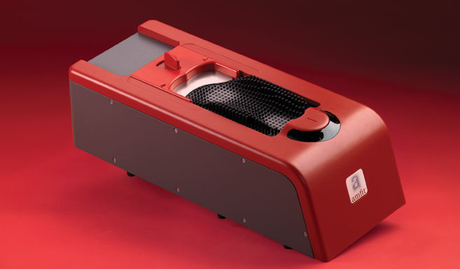
The Amfit Contact Digitizer is a pin-contact scanner: 538 pins physically deflect soft tissue under operator-controlled pressure to reach the bony landmarks underneath. It's a different category of capture from photo or laser systems, which read the soft-tissue surface. Neither approach is universally “better” in the literature; they measure different things and produce different inputs to the prescription.
What the pin-contact approach gives a practice specifically is operator control. The provider or staff member running the scan can adjust pressure for the patient on the table: lighter for diabetic or geriatric patients, firmer for athletes. The scan that flows into Correct & Confirm is the bony-architecture scan you actually want to build the orthotic on, not just what the skin looked like at rest.
Whether that matters for your practice comes down to volume and clinical philosophy. For a practice doing 30+ pairs a month and treating sports, work-comp, and complex postural cases, the difference between “good enough” capture and millimeter-grade capture shows up in remake rate and patient-reported fit over a six-month window. For a lower-volume practice doing primarily comfort orthotics, the case is softer.
A defensible exam-room workflow
What does an integrated workflow look like in practice? Roughly this:
- Visual screen. Standard postural and gait observation. Note asymmetries, obvious pathology, and patient-reported pain pattern.
- Static foot capture. Patient on the scanner. Two minutes. Pin-contact (or whichever capture method you've standardized) produces the digital cast.
- Clinical review. Software opens the cast against your prescription. You confirm metatarsal pad placement, posting, heel skive, and any condition-specific accommodations.
- Prescription locks before fabrication. Whether you're milling in-clinic or sending to central fab, the design is reviewed before EVA gets cut.
- Outcome tracking. Re-exam at 4–6 weeks. If fit is off, the digital cast is preserved: you adjust on the file, not from scratch.
The point isn't to replace clinical judgment with technology. It's to give the clinical judgment something solid to ride on.
Bottom line
Visual gait analysis remains a useful screen. The literature does not support treating it as a measurement-grade input on its own. Layering an instrumented foot capture onto the exam (pin-contact, laser, or otherwise) gives the orthotic prescription a defensible foundation, and gives the practice something to point at when fit is questioned six months later.
The honest read of the research: scanners are not magic, but they are reproducible. That's the property the prescription was missing.
References
- Reliability and Validity of Observational Gait Analysis by Physical Therapists. J Phys Ther Sci, 2025. PMC12445358
- Measurement properties of observational gait analysis in patients with stroke: a systematic review. PMC, 2024. PMC12153243
- Does Scanner Choice Matter for the Design of Foot Orthosis? Sensors, 2025. MDPI 25/3/869
- Comparison of 3D scanning versus traditional methods of capturing foot and ankle morphology for the fabrication of orthoses: a systematic review. J Foot Ankle Res, 2020. JFAR 13:48